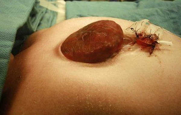
Ectopia Cordis Definition
Ectopia cordis is a Latin word, which literally means “outside” and “heart”. It is a rather rare congenital malformation that concerns the heart’s abnormal location. Normally, the heart is located in the center of the chest cavity where it resides in the space between the lungs (inferior mediastinum) and lies on the diaphragm with its more pointed apex directed to the left side.
Moreover, the heart is approximately located in the fifth intercostal space. However, in Ectopia cordis case, the heart is abnormally located either partially or completely outside of the chest cavity. The estimated prevalence rate of ectopia cordis is 0.079/10,000 births and is said to occur more in female infants.
Causes Of Ectopia Cordis
Although the exact, precise etiology or cause of the disease is still not known, Ectopia cordis is largely attributed to the improper development of the chest cavity’s structure during the embryonic stage (specifically in the 8th day of the embryonic life). The midline mesoderm unsuccessfuly follows the proper maturation it was intended to have and the anterior (ventral) body wall formation fails to develop properly.
Basically, what happens is, the lateral body walls, which are mainly responsible for the proper union or connection of the ventral or anterior walls, have failed to initiate fusion. Usually, this improper or incomplete fusion of the ventral walls occurs in the 9th week of life.
In addition, there is also a mechanical theory proposed to explain ectopia cordis. This mechanical theory is the early rupture of chorion and/or yolk sac. The chorion and/or yolk sac causes compression of the thorax that consequently puts a stop to the midline fusion. Furthermore, the amniotic band syndrome is also being proposed as a mechanical cause of ectopia cordis.
Amniotic band syndrome otherwise called as amniotic band sequence is generally a group of birth malformations caused by the ensnarement of the infant anatomical parts (frequently the upper or lower extremity) in amniotic strips whilst still in the uteros that leads to malformation, disruption or deformation. In effect, the defective ventral wall is now not capable of providing the heart its necessary shield of protection. The heart is then rendered unprotected by its supposed to be natural protective structures, which are the pericardium, sternum and even the skin.
Moreover, the disease can occur as a solely isolated malposition of the heart or it can be also linked to a much larger case of anterior body wall defects that involves the abdomen, thorax or both. The most common thoracic birth defects associated with ectopia cordis are tetralogy of fallot, atrial septal defect, tricuspid atresia, double outlet right ventricle and ventricular septal defect.
On the other hand, the most common abdominal birth defect associated with ectopia cordis is omphalocele (a congenital malformation wherein the infant’s intestine, liver and/or other abdominal organs stick out of the belly button or navel). Furthermore, ectopic cordis has also been associated with chromosomal abnormalities like trisomy 18 and Turner syndrome. Cleft lip and cleft palate are the most common facial defects.
Ectopia Cordis Pictures

Picture 1 : Ectopia cordis Image

Picture 2 : Ectopia cordis
Types of Ectopia cordis
Ectopia cordis is medically classified into four categories depending on the position of the heart outside the chest cavity. These categories are as follows:
- Cervical Ectopia cordis – This type of ectopia cordis constitutes 5% of the total cases. The exposed heart is located in the neck of the infant.
- Thoracic Ectopia cordis – This type of ectopia cordis constitutes 65% of the total cases. The exposed heart is located in the anterior of the sternum.
- Thoracoabdominal ectopia cordis – This type of ectopia cordis constitutes 20% of the total cases. The heart is located in the area between the thorax and the abdomen.
- Abdominal ectopia cordis – This type of ectopia cordis constitutes 10% of the total cases. The heart is located in the abdomen of the infant.
Among the four categories, the thoracic and thoracoabdominal ectopia cordis are predominantly the most common type.
Ectopia cordis association with Cantrell’s pentalogy
Pentalogy of Cantrell, also known as thoraco-abdominal syndrome is a rare and fatal congenital anomaly that involves the diaphragm, pericardium, heart, lower sternum and the abdominal wall. It has a prevalence rate of 1 per 100,000 births and is generally more common in male infants.
Ectopia cordis is one of the five characteristic presentations of the Pentalogy of Cantrell, along with omphalocele, anterior diaphragmatic hernia, sternal cleft and intracardiac defects such as ventricular septal defect and diverticulum of the left ventricle.
Ectopia Cordis Treatment and Repair
More often than not, therapeutic abortion or pregnancy termination prior to age of viability is almost always being considered for the parents, upon discussion with the physician. However, if termination is out of the equation, careful and deliberate search for associated abnormalities and cautious chromosomal analysis should be employed to determine the severity of the condition. Moreover, due to the disease’s very poor prognosis a non-aggressive approach is recommended.
As to the treatment regimen for ectopia cordis, there have been limited treatment options due to the uncommonness of the condition and the speedy mortality of the infant shortly after delivery. Although there have been successful surgical interventions for ectopia cordis, the mortality rate is still very high.
One of the surgical efforts that are being employed is the instantaneous covering of the naked heart and abdominal contents with silastic prosthesis. If there is a presence of associated intracardiac defects, then this is being attended and corrected first, prior to the replacement of abdominal contents. If the infant survived this sugical operation then he/ she may be viable for orthotopic heart transplantation.
Prognosis and Survival
The prognosis of Ectopia cordis generally depends on three factors, which are as follows:
- Location of the defect
- The extent of intracardiac defects; and
- If there are associated abnormalities present.
In most cases, the prognosis is generally poor. Most babies are stillborn while others expire shortly after delivery (within hours up to a few days of life) due to hypoxemia, cardiac failure and infection. Efforts for surgical intervention have been rendered difficult and generally unsuccessful due to complications brought about by associated anomalies.
References:
http://en.wikipedia.org/wiki/
http://www.ncbi.nlm.nih.gov/
http://www.medicinenet.com/

