What is Ectrodactyly?
The term, ectrodactyly comes from the Greek word “ektroma”, meaning abortion and “daktylos”, meaning finger. Essentially, ectrodactyly literally translates to the abortion of a finger.
In the medical field however, ectrodactyly is otherwise known as split hand/split foot malformation or SHFM. It is a relatively rare congenital limb malformation wherein there is absence or deficiency of one or more central fingers or toes in the hand or foot. This central absence of fingers or toes is mainly because of the absence of the central rays. Moreover, the condition is also accompanied by fusion of the remaining peripheral digits.
There may also be instances where metacarpals, metatarsals and phalanges are affected with aplasia/hypoplasia. Due to these circumstances, the appearance of ectrodactyly very much resembles that of a lobster’s claw that is why the condition is also widely dubbed as the “lobster claw syndrome”. Although the “lobster claw syndrome” is not widely used due to the fact, that it may cause unintentional disrespect towards the patient.
Furthermore, the prevalence rate of the condition is around one per 90,000 births. In addition, there is no definite record of partiality as to which gender (male or female) is more affected.
Ectrodactyly Causes and Genetics
Ectrodactyly may present as a single birth defect (non-syndromic form) or it may also present as one of a group of birth defects that is present in an infant upon birth (syndromic form). For instance, it may be part of a certain syndrome known as Ectrodactyly–ectodermal dysplasia–cleft syndrome or EEC, which will be further discussed in a separation section below.
Ectrodactyly in EEC may have a significant effect on one’s health condition; however, in cases where ectrodactyly is only a single birth defect, it rarely presents as a significant health risk.
Anyhow, ectrodactyly is said to be caused by a hereditary gene mutation. Since it is hereditary, there is a high chance of a parent with ectrodactyly to pass on the trait to his/her offspring. Usually, the infant inherits the condition through autosomal dominant mode with associated reduced penetrance. However, X-linked and autosomal types may also occur but only in seldom cases.
On the other hand, the gene mutation or chromosomal rearrangement in ectrodactyly is either in the form of translocation or duplication and deletion, primarily in chromosome number 7. This chromosome number 7 houses the two homeobox genes known as DLX5 and DLX6 respectively. An alteration in chromosome number 7 (either a translocation or deletion) may result to associated loss in hearing. Moreover, ectrodactyly type 1 is the only ectrodactyly linked to sensorineural hearing loss.
In a very detailed research done on mouse models, a certain entity, the apical ectodermal ridge, effectively shortened as AER is vital in the discussion of ectrodactyly occurrence. This apical ectodermal ridge is essentially located at the tip of the developing finger or toe bud. Fundamentally, the main cause of ectrodactyly is attributed to the disturbance or interruption in the AER function.
Either of the following may disturb the AER function: genetic defects and extrinsic factors such as caffeine, ethanol, methotrexate, cocaine, cadmium and valproic acid among others. Basically, the apical ectodermal ridge, along with the progress zone and the zone of polarizing activity form the three specific cell collection that are responsible for yielding signalling molecules for the proper formation of distal limbs. Without these collective factors, proper finger and toe formation is unattainable.
However, the specific genetic cause of human ectrodactyly is difficult to pinpoint due to a number of factors such as the following:
- the inadequate quantity of families associated with split hand/split foot malformation
- the hefty amount of morphogens associated or included in growth of the extremities
- the occurrence of complex interaction between the said morphogens; and
- the pre-deduced paticipation of several gene and widely covering regulatory factors in several cases of SHFM
The only genetic mutation associated with human entrodactyly is the one that happens in the TP63 gene.
Ectrodactyly Pictures

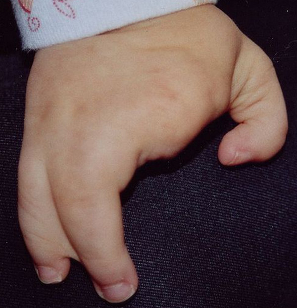
Picture 1 : In 1 year old child – Ectrodactyly on the hand

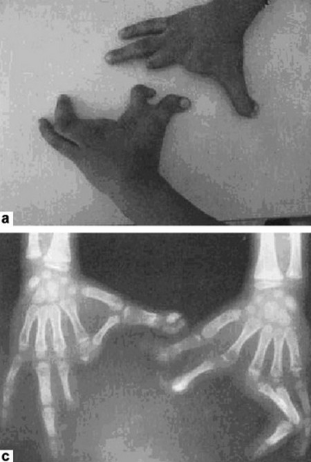
Picture 2 : Ectrodactyly – Split hand feet malformation – median clefts of the hands, hypoplasia/ or aplasia of the phalanges, and metacarpals.

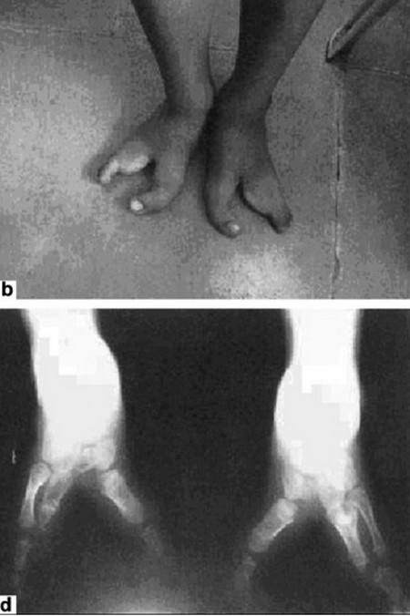
Picture 3 : Ectrodactyly – Split hand feet malformation – median clefts of the feet, hypoplasia/ or aplasia of the phalanges and metatarsals
Diagnosis of Ectrodactyly
The diagnosis of ectrodactyly may entail some difficulties and complications due to the presence of overlapping symptoms with other ectodermal dysplasia syndromes. Although diagnosis may sometimes be complex, it is however, very possible to do correct diagnosis. Correct diagnosis may be possible through the use of the following diagnostic exams: X-ray, skin biopsy, kidney imaging test and DNA testing among others. These diagnostic exams may be used in combination for appropriate diagnosis.
Activities of daily living in people with ectrodactyly
People affected with ectrodactyly as a single birth defect are mostly nearly as functional as normal people are. They can even perform tasks requiring skillful manipulation with no or little difficulties. Tasks involving the hands such as writing and personal grooming are done aptly. Although the condition may affect the performance of the activities of daily living in a minute way, the end result of those tasks however are more often than not, satisfactory.
Moreover, people with ectrodactyly have stable jobs of their own and some even opted to raise their own families.
Associated conditions or syndromes with ectrodactyly
Ectrodactyly is more often than not associated with a number of genetic defects, some of which will be each discussed in the following:
- Ectrodactyly – ectodermal dysplasia – cleft syndrome (EEC) – EEC is the most common associated birth defect with ectrodactyly. It is caused by an alteration in the TP63 gene, which is usually a missense mutation. As may be inferred from the name itself, the condition is typified by a triad of distinct physical attributes, which are ectrodactyly, ectodermal dysplasia and of course, cleft lip or palate. EEC mutation occurs in the DNA binding domain of p63. Moreover, the associated cleft lip or palate in EEC is usually managed through surgical intervention. On the other hand, the ectodermal dysplasia or the absence of sweat glands that results to dry, scaly skin and scalp is managed through frequent intake of cold liquids to maintain thermoregulation as well as hydration.
- Ectrodactyly – Cleft Palate (ECP) syndrome – As the name suggests, ECP is typified by a physical presentation of ectrodactyly and cleft palate. Its mode of transmission is usually autosomal dominant.
- Ectrodactyly – Ectodermal Dysplasia – Macular Dystrophy (EEM) syndrome – EEM is an autosomal recessive disorder. Moreover, the macular dystrophy in EEM is a progressive disease of the eye. Mascular dystrophy occurs due to the alterations in CDH3 found in human chromosome 16. Aside from ectrodactyly, ectodermal dysplasia and macular dystrophy, EEM is also characterized by the following characteristics: webbed fingers or toes that are medically referred to as syndactyly, hair loss or hypotrichosis and dental problems.
- Ectrodactyly – Fibular Aplasia/Hypoplasia (EFA) syndrome – EFA’s mode of transmission is usually autosomal dominant with reduced penetrance. It is usually typified by a number of physical characteristics such as fibular deformity, split hands and short fingers among others.
- Ectrodactyly – Polydactyly – In EP, ectrodactyly has an accompnaying presence of polydactyly, which is basically the presence of an extra digit. Polydactyly occurs in an autosomal dominant fashion in a single gene.
Medical and Surgical Management
Ectrodactyly is more often than not, managed through surgical intervention. Surgical intervention may help in improving appearance as well as function of the hands or feet in activities of daily living, most especially. However, this may not entail that the hands or feet function exactly the way, as regularly formed hands will. Furthermore, surgical repair of associated symptoms such as cleft lip or palate may also be performed. Individuals who undergo surgical repair of the lip and palate may require lengthy physical as well as speech therapies to ensure better recuperation and functioning.
Moreover, the absence of sweat glands may cause the integumentary part of the body to be brittle, which may in turn present further problems. That is why medical intervention should also be done for this associated symptom to prevent future problems.
In addition, employment of early occupational and physical therapies may greatly help people with ectrodactyly to do simple activities of daily living such as writing, eating, grooming and picking things up among others. Moreover, in other cases, some may opt to using prosthetics, which may also be highly beneficial to the patient.
Since the condition is genetic or hereditary in nature, the parents should be counseled for possible reappearance of the condition in future children. In this case, the physician usually offers to conduct an antenatal diagnosis through ultrasonography.
Prognosis for Ectrodactyly
The prognosis of people with ectrodactyly, especially those who have ectrodactyly as a single birth defect is very good. The condition rarely present as a significant health risk to the one affected, except of course for minor difficulties. The life expectancy may range from very slightly decreased to normal.
However, in people with syndromic form of ectrodactyly, a number of health risks and problems may occur such as dealing with the individual’s sweating problems. However, the prognosis is still very good, with a life expectancy of slightly reduced to normal.
Other species affected with ectrodactyly
The occurrence of ectrodactyly or SHFM is not only limited to humans. Other living organisms that may also be affected with this condition are cats, dogs, frogs and toads, chickens, cows, rabbits, marmosets, mice, salamanders and West Indian manatees.
References:
http://emedicine.medscape.com/
http://www.ncbi.nlm.nih.gov/


